Illuminating tumour micro environments
Advanced technical imaging capabilities and research projects supported by MWC are helping to identify new prognostic and diagnostic biomarkers to advance potential therapeutic targets for many different cancers, narrowing the gap between laboratory discoveries and clinical applications.
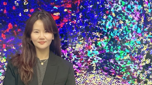
Dr Saem Park with an image of the tumour microenvironment of melanoma-infiltrated lymph node tissue, visualised by multiplex immunofluorescence using the Vectra Polaris imaging system.
Dr Saem Park, research fellow from the University of Auckland and an internationally recognised leader in tissue imaging technologies, was appointed as one of three research facilitators within the MWC cancer theme for 2023/2024.
In this MWC-funded role, Dr Park has supported projects from 15 MWC-associated research groups across New Zealand. As well as applying her advanced imaging expertise to projects, she has mentored other researchers and facilitated the adoption of new advanced tissue imaging techniques.
Dr Park developed study protocols and designed multicolour antibody panels for tissue imaging and guided image analysis workflow for various human tissues, in research areas including endometriosis, tumour hypoxia, melanoma, liver, pancreatic, colorectal, ovarian and cervical cancers as well as in mouse cancer models. Furthermore, she established an analysis pipeline for comprehensive in-depth tumour immune phenotyping.
Under her leadership, MWC researchers began trialing the Vectra Polaris tissue imaging system, a new single-cell analysis workflow technology using formalin-fixed paraffin-embedded tissue samples alongside the Visium High Definition spatial gene expression assay from 10X Genomics (a next-generation molecular profiling solution for classifying tissue based on total mRNA).
The new automated process uses a high throughput, multi-colour digital tissue imaging platform (Vectra Polaris) which loads up to 80 slides stained in seven different colours. An entire tissue section can be scanned while simultaneously digitally quantifying different cell types and assessing their potential interactions using spatial approaches.
The new system and Dr Park’s expert guidance is significantly extending MWC’s ability to analyze tissue samples, more than tripling output from earlier years.
Dr Park says “With the shared knowledge of the new imaging workflow, MWC research groups have accelerated digital tissue profiling on melanoma, lung, colorectal, breast, head and neck, ovarian and liver cancers.
“In turn, this work is advancing the identification of more reliable predictive biomarkers for cancer immunotherapy.”
These skills and expertise around imaging technologies have also helped MWC researchers to progress new and international collaborations with the University of Oxford (United Kingdom), the Walter and Eliza Hall Institute of Medical Research (Australia) and Korea University Guro Hospital (South Korea), conference presentations, and grant applications.
“Overall,” says Dr Park, “the project has laid a foundation for sustained progress in tissue imaging and cancer research, allowing MWC researchers to tackle complex questions with advanced spatial biology tools.”