New imaging improves our understanding of the immune system
Maurice Wilkins Centre researchers have used novel imaging technology to reveal the structure and inner workings of entire lymph nodes, core components of the body’s immune system.
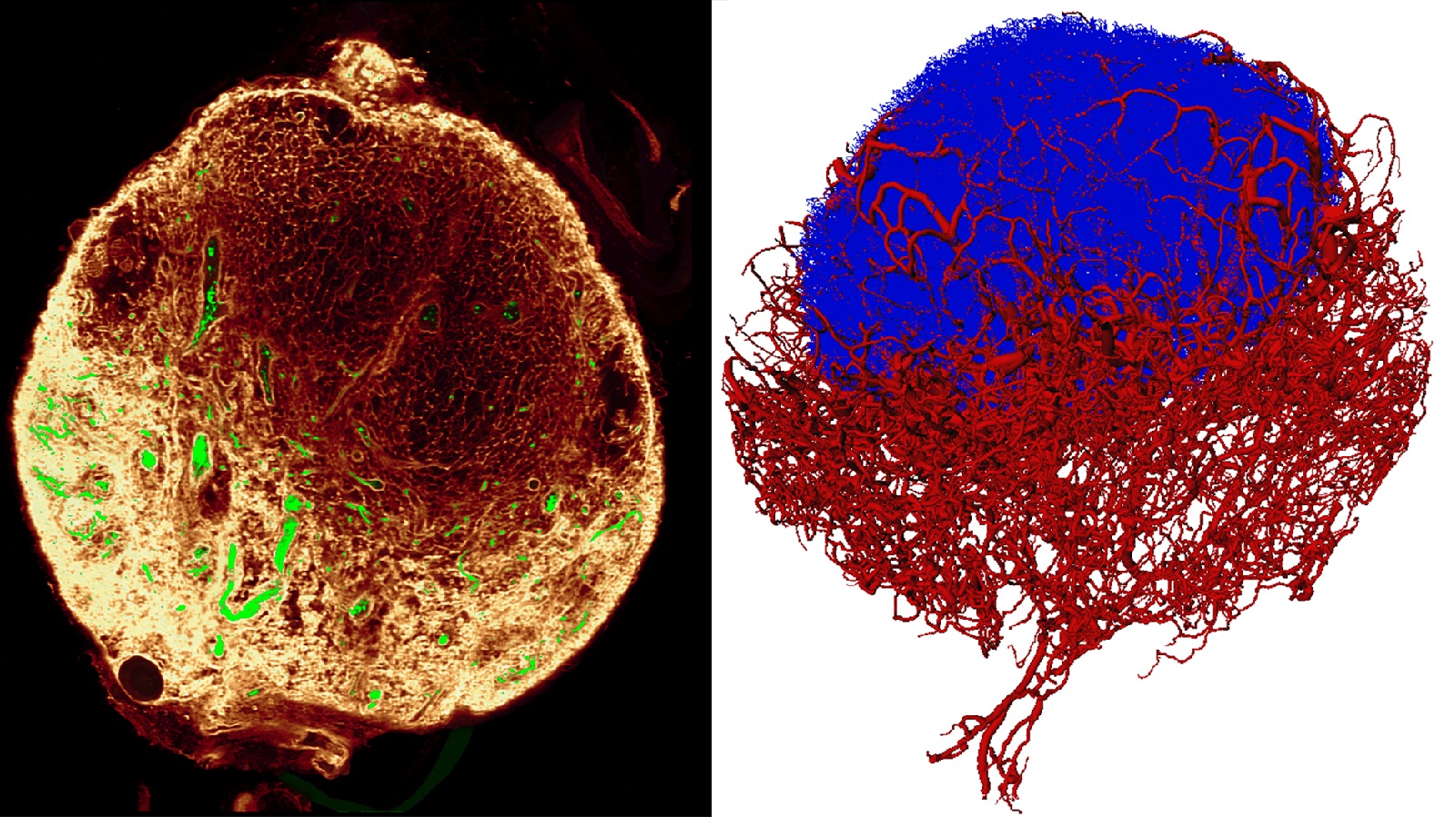
MWC-funded research by Dr Inken Kelch and a team of Auckland scientists led by Prof Rod Dunbar, which involved the use of novel 3D imaging system to map out an entire mouse lymph node, has been published in the journal PLOS Biology.
Developed by collaborators at the Auckland Bioengineering Institute (ABI), Dr Gregory Sands and Associate Prof Ian LeGrice, this New Zealand technology features a customised confocal microscope capable of producing very high resolution images of large areas of tissue.
Novel tech allows high resolution imaging of a relatively large area
"The problem with conventional imaging methods is that the higher the resolution you seek, the smaller the area that you can cover. There's usually a trade-off between size and resolution,” explained Dr Kelch.
“Our collaborators built a system where you can achieve high resolution imaging but you can also cover a large area. This was ideal for what we wanted – to look at lymph nodes, which are small organs with a very intricate, complicated architecture.”
Illustrated in their paper, the powerful 3D images that Dr Kelch and her co-authors including Prof Anthony Phillips were able to take using the new system provide a detailed ‘big picture’ of a lymph node dissected from a mouse. They reveal how various regions of the lymph node are supplied by a fine tubing system called ‘conduits’, including their close relationship with blood vessels and their anatomy in regions where immune responses are kick-started.
The architecture of the lymph node supports crucial interactions between immune cells, said Dr Kelch. “We often talk about lymph nodes as being the headquarters of the immune system where immune responses are formed and different immune cells can come together and talk to each other. It is now becoming clear that the structural framework of the lymph node is needed to support and shape these interactions.”
To get the most of their unique images, Dr Gib Bogle developed software to turn the conduit network into a computer reconstruction, enabling an extensive range of measurements and in particular modelling of the conduit anatomy’s roles in lymph node function.
Better understanding of lymph node biology and function may guide cancer therapy research
“We’re particularly interested in the homing zone for T cells,” added Dr Kelch. “T cells are important for immune responses to infections and cancer, they are the acting agents – the ‘police force’ – that can seek and destroy a tumour. Interestingly, T cells are highly motile and constantly scan lymph nodes for alarm signals. We are now able to simulate their migration in a realistic context using our conduit map.
“A large amount of research is being carried out into the role of T cells and how we might be able to manipulate them in a way that is advantageous for patients with cancer in particular. However, the normal biology and function of lymph nodes and the immune system as a whole is yet to be fully understood. This is why our work is important – it will provide researchers with a better insight into how lymph nodes are organised to support immune function and gives us a new comprehensive map to aid modelling of the immune system."
Reference:
Kelch ID, Bogle G, Sands GB, Phillips ARJ, LeGrice IJ, Dunbar PR. High-resolution 3D imaging and topological mapping of the lymph node conduit system. PLoS Biol 2019:17(12): e3000486, DOI: https://doi.org/10.1371/journal.pbio.3000486