Advanced technology leads to discovery of a new cell in human skin (2014)
MWC scientists have developed new methods to identify the different types of cells in human tissues – including very rare ones.
 Their latest exciting research led to a cover article in the journal Stem Cells and Development that is already being cited in a host of leading international publications.
MWC Research Fellow Dr Anna Brooks, working in the laboratory of MWC Director Professor Rod Dunbar, has pioneered analysis of the cells in human tissues using a technique called flow cytometry. Part of her work has focussed on the cells that make the connective tissues holding the body together. Dr Vaughan Feisst in Rod’s lab has been studying the stem cells that give rise to these cells and, using Anna’s flow cytometry techniques, the team went looking for such stem cells in human skin.
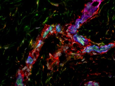
Flow cytometry allowed the team to first identify these rare cells in human skin samples and then to determine molecules that could be used to locate them. Working with the lab’s microscopy expert Jenni Chen, they discovered these cells are located on the edge of blood vessels, just as they are in some other human tissues.
“This discovery is pretty fundamental. It opens the door to advances in both skin healing and treatment of skin diseases where we suspect these cells malfunction,” Rod says.
The team is now working with Dr Michelle Locke, Senior Lecturer in Plastic and Reconstructive Surgery at the University of Auckland, to understand how this knowledge can be used to improve treatment of skin injury and the impaired healing that many surgical patients suffer. Plastic surgeons and their patients at Middlemore Hospital in Auckland have also been crucial in generously donating tissue for the research.
“This is a good example of the new type of work that the MWC has enabled,” says Rod. “The investment in technical capability over many years has led to unique methods that generate exciting new research results. And the interface between bench scientists and clinicians that the MWC has fostered ensure the work stays highly relevant to patient care.”
The team began analysing the cells in human skin to understand how to design better vaccines for use in immune therapy for cancer patients. While analysing the immune cells in human skin, the team realised they could apply similar techniques to other medical problems.
Image: cutting through human skin reveals the stem cell niche, around the walls of blood vessels. The cells that form the walls of blood vessels, pericytes, stain positive for pericyte marker CD146 (Blue), and are surrounded by cells that stain for mesenchymal cell marker CD90 (Red). Some of these cells are also positive for the marker CD34 (Green), and it is these cells, that appear yellow due to the combination of CD90 and CD34 (Red and Green), that are the stem cells. Image courtesy of Vaughan Feisst.